
First, a word about normal aging
One fact matters more than any single term on the report. Many spine MRI findings show up in people who have no pain at all. In a well-known 1994 study in the New England Journal of Medicine, researchers scanned the low backs of 98 adults without back pain. About half had a disc bulge at one level or more, and about a quarter had a disc protrusion.
A later review of more than 3,000 pain-free people found the same thing across age groups. Disc degeneration showed up in roughly a third of 20-year-olds and in most people over 60. Disc bulges followed a similar climb with age.
So a report full of findings does not always mean something is wrong. The question a spine surgeon asks is whether the pictures match your symptoms and your exam. When they match, the MRI is very helpful. When they do not, the findings may simply be part of normal aging.
How the report is laid out
Most reports list each spinal level from top to bottom. Levels are named by the two bones on either side of the disc. L4-L5 is the disc between the fourth and fifth lumbar vertebrae in the low back. L5-S1 is the lowest lumbar disc, just above the sacrum. C5-C6 is a disc in the middle of the neck. In the low back, most disc problems happen at L4-L5 and L5-S1. In the neck, C5-C6 and C6-C7 are the most common levels.
The Impression section at the end is the radiologist's summary. It is the part most doctors read first.
Disc terms
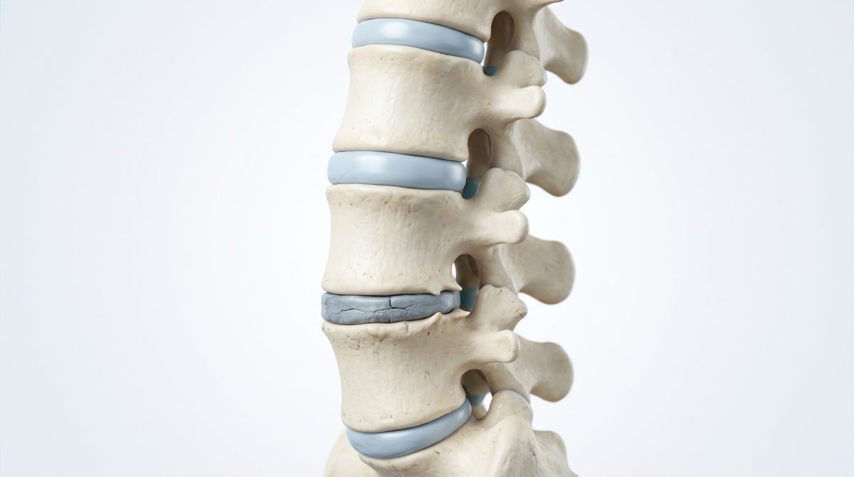
Disc degeneration or desiccation. The disc has lost some of its water and looks darker on the scan. This is part of aging and is sometimes called degenerative disc disease, even though it is not really a disease. It is often painless.
Loss of disc height. The disc is thinner than the ones around it. This can narrow the openings where nerves exit.
Disc bulge. The disc spreads out evenly beyond the edges of the bones, a bit like a hamburger patty that is wider than its bun. Bulges are very common and are often not a cause of pain.
Protrusion, extrusion and sequestration. These describe a herniated disc, where part of the soft inner disc pushes out through the tough outer ring. A protrusion is a smaller, broad-based push. An extrusion pushes out further, with a narrow neck. A sequestered or free fragment has broken off completely. A larger herniation is more likely to press on a nerve, but size alone does not predict pain.
Annular fissure or annular tear. A small crack in the outer ring of the disc. The word "tear" sounds alarming, but these are common in people without pain and do not mean an injury happened.
Modic changes. Changes in the bone right next to a disc, graded as type 1, 2 or 3. Some research links type 1 changes with low back pain, but the meaning is still debated.
Narrowing and nerve terms
Stenosis. Stenosis simply means narrowing. Central canal stenosis is narrowing of the main tunnel that holds the spinal cord or nerve bundle. It is often graded as mild, moderate or severe. In the low back, it can cause leg pain and heaviness with walking, a condition called lumbar spinal stenosis.
Foraminal stenosis or foraminal narrowing. Narrowing of the side opening, the foramen, where a nerve root leaves the spine. It can pinch that single nerve and cause pain down one arm or one leg.
Lateral recess stenosis. Narrowing in the gutter just before the foramen, where the nerve root travels on its way out.
Nerve root impingement, contact or compression. Something, often disc or bone spur, is touching or pressing on a nerve root. "Contact" is milder than "compression". This finding matters most when your symptoms follow that same nerve.
Cord compression or myelomalacia. These are neck and upper back terms. Cord compression means the spinal cord itself is being pressed. Myelomalacia means the cord shows signal changes from that pressure. These findings deserve a prompt visit with a spine surgeon, even if your pain is mild, because the cord does not heal as easily as a nerve root.
Joint, bone and alignment terms
Facet arthropathy or facet hypertrophy. Arthritis of the small paired joints at the back of each level. The joints can enlarge and add to narrowing. Mild facet arthritis is common with age.
Ligamentum flavum thickening. A ligament at the back of the canal has thickened or buckled. Together with a bulging disc and enlarged facets, this is a common cause of stenosis.
Osteophytes or bone spurs. Small bony growths at the edges of the bones or joints. They form over years and are sometimes called spondylosis.
Spondylolisthesis or listhesis. One vertebra has slipped forward (anterolisthesis) or backward (retrolisthesis) on the one below. It is graded by how far it has slipped.
Scoliosis or curvature. A sideways curve. An MRI shows it, but standing X-rays are the most reliable way to measure it. Our guide to adult scoliosis has more.
Straightening of the normal lordosis. The gentle inward curve of the neck or low back looks flatter. It is often due to muscle spasm or the way you were lying in the scanner and is usually not meaningful on its own.
Other phrases you may see
- "Clinical correlation recommended" is the radiologist asking your doctor to compare the images with your symptoms and exam. It is a standard phrase, not a hidden warning.
- "Unremarkable" means normal.
- "Incidental finding" means something noticed by chance that is not related to why the scan was done, such as a small kidney cyst. Ask your primary care doctor whether it needs follow-up.
- "Post-surgical changes" refers to scar tissue or hardware from a prior operation.
When to call us
If your report mentions cord compression, myelomalacia or severe stenosis, or if the findings line up with pain, numbness or weakness down an arm or leg, it is worth having a spine surgeon review the images with you. Bring the disc or image link along with the written report, because the pictures often tell more than the words.
Common questions
My report says "herniated disc." Do I need surgery?
Usually not. Many herniated discs shrink over weeks to months, and most people improve with time and nonsurgical care. Surgery may be considered when leg or arm pain stays severe for weeks, or when weakness is present or getting worse.
Why does my report sound so much worse than I feel?
Radiologists list everything they see, including common age-related changes. Many of those findings cause no symptoms. Your doctor's job is to sort out which findings, if any, explain your pain.
Is an MRI better than an X-ray for back pain?
They show different things. An MRI shows discs, nerves and soft tissue. Standing X-rays show alignment, slippage and how the spine behaves under your body weight. Surgeons often want both before planning an operation.
Can I send my MRI before the visit?
Call the office first. Our staff can tell you how to get the images to us so the surgeon can look at them during your appointment. Please do not send images or medical details through the website form.
Sources
- Jensen MC, et al. Magnetic resonance imaging of the lumbar spine in people without back pain. N Engl J Med. 1994.
- Brinjikji W, et al. Systematic literature review of imaging features of spinal degeneration in asymptomatic populations. AJNR Am J Neuroradiol. 2015.
- Lumbar MRI scan, MedlinePlus Medical Encyclopedia (NIH)
- Spinal stenosis, MedlinePlus Medical Encyclopedia (NIH)